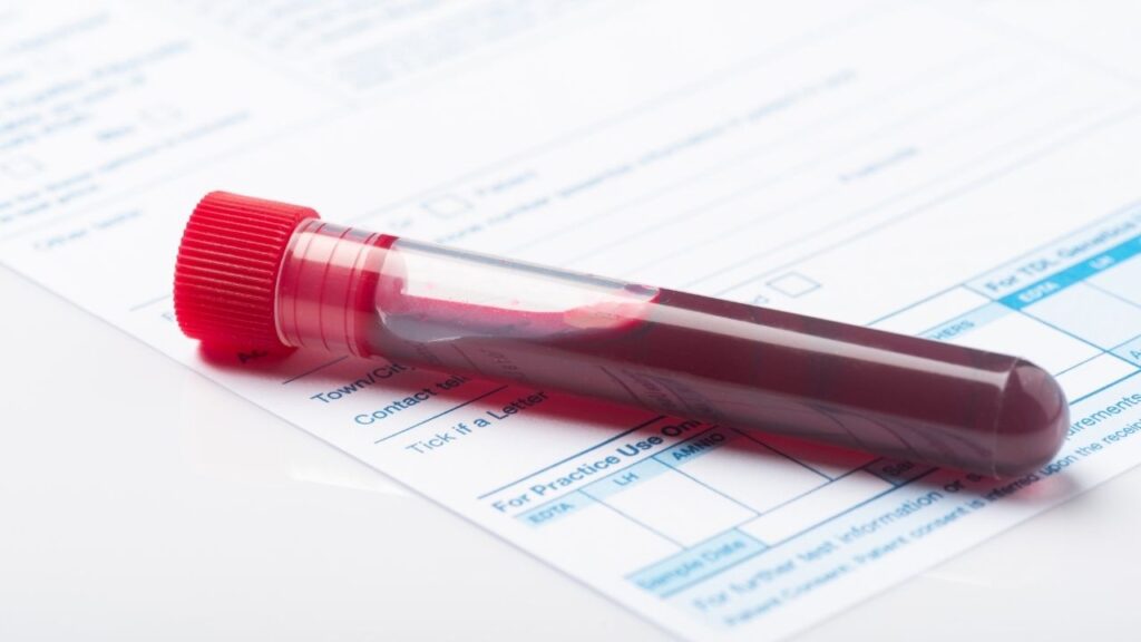
A biopsy for dog cancer is a reliable and accurate method for getting a tumor type, how aggressive it is, and whether it is malignant. Biopsy can be the key to early detection and will help determine the prognosis and course of treatment.
Key Takeaways
- Biopsies vary in cost, depending upon the location, the difficulty, and the size of the sample being taken. A simple punch biopsy at your general practitioner’s hospital might cost $350, while a more invasive biopsy performed by a board-certified surgeon could cost $2,500 or more.
- Your dog may spend a few hours at the hospital for a biopsy, and perhaps longer if anesthesia is needed. The biopsy report from the lab can take a week or even ten days to get back.
- Dogs recover well from biopsies in general, depending upon the location and how involved the procedure was. Dogs usually go home the same day. Healing time varies, with skin biopsies healing quicker than abdominal biopsies. If sutures are involved, they are usually removed a week or two weeks after the biopsy.
- Dogs usually are put under anesthesia for a biopsy in deep areas of the body. If they need a biopsy of the skin or surface, they may only require sedation to make sure they are still and comfortable.
What is a Biopsy for Dog Cancer?
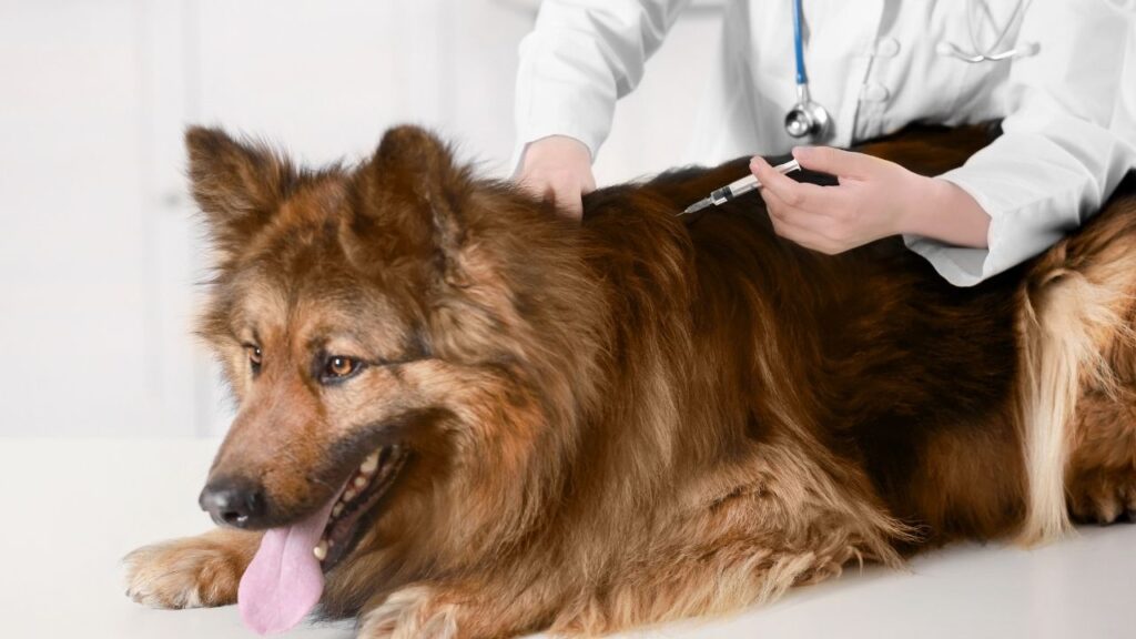
A biopsy is a procedure to surgically remove a sample of tissue from your dog to send to the laboratory for more information.
In many cases, it’s the most accurate way to diagnose cancer and get a prognosis. Prognosis is the likely outcome and the chance of successful treatment or recurrence of your dog’s cancer.
What Happens During a Biopsy for Dogs
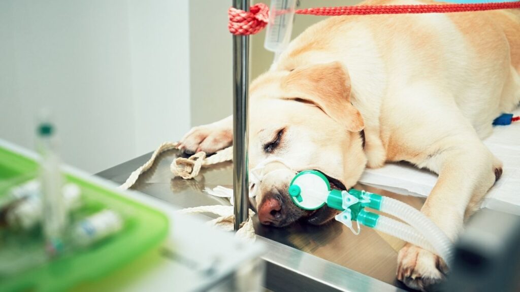
When a sample of tissue or tumor is surgically removed from your dog, he will need numbing, sedation, or general anesthesia. Your veterinarian will decide which of these is best, depending on the tumor or affected tissue size, location, and your goals.
Sometimes biopsies are performed as an add-on procedure during other surgeries. For example, dogs who get dental cleanings might get a biopsy if the veterinarian finds something suspicious during the cleaning. Samples may also be taken during abdominal exploratory surgery, endoscopy, or ultrasound depending on diagnostic goals, your dog’s health in general, and the location and size of the tumor.
Once the tissue sample is removed, it is placed in a special preservation solution and sent to a laboratory for evaluation.
Typically, once at the laboratory, the sample is cut into very small pieces and a pathologist (diagnostic specialist) reviews the samples under a microscope.
After review, a diagnosis is made, and the pathologist writes a detailed report. This report is sent to your veterinarian to share with you as they propose a plan for treating your dog’s specific cancer.
The whole process usually takes a week or two, depending on the lab and how many samples are being processed at a the time.
Where to Get a Biopsy for Dog Cancer
General practice veterinarians commonly perform many different types of biopsies.
However, there are cases where it is in your dog’s best interest to go to a specialist – usually an oncologist or surgeon. This is especially true if advanced imaging is needed to get the biopsy samples or if any treatment may take place at the same time.
If your general practice doctor is not comfortable or feels that your dog would benefit from specialty referral, they will provide you with these options.
The samples themselves are always sent to a laboratory pathologist for review. This is usually in the mail, unless there is an on-site lab.
Why Does Your Dog Need a Biopsy?
Biopsy allows for many sections of tissue to be prepared in different ways and to look at a large sample of cells. With so many cells to look at, in the arrangement that they are in your dog, the pathologist can get specific information to help your veterinarian know how to treat your dog’s cancer.
Biopsy is used to:
- Differentiate between malignant (cancerous) and benign tumors
- Determine what type of tumor it is, and if it was all removed
- Get more specific answers when cytology (looking at a fine needle aspirate sample under a microscope) alone is not helpful enough
The biopsy results help your veterinarian create a treatment plan and to estimate how successful it may be.
Biopsy Types for Dogs with Cancer
There are several ways to get a biopsy sample. Your veterinarians will consider where the tumor is located, how many blood vessels are in that area, the risk to your dog, and what your ultimate goals are before selecting a sampling method.1
Core Needle Biopsy (CNB)
Core needle biopsy, also known as a core biopsy, requires a needle with cutting edges and a hollow core. This is inserted into the affected tissue to get a sample.
- There are different needle sizes that yield different biopsy sizes/widths depending on location, risk of bleeding, and other factors.
- Sometimes, the sampled area will be closed with sutures or staples, but this is not always needed.
- Often the area will be numbed, and sometimes sedation is needed for safety, to keep the patient still, and to get the best possible samples.
- Full general anesthesia may be needed to safely sample certain organs.
- Sometimes imaging methods such as ultrasound or CT may be used to guide the veterinarian getting the sample.
Core needle biopsy is commonly used for skin masses, masses of internal organs, and bone tumors.
Your dog may already have gotten a fine needle aspirate, which is similar to this type of biopsy, but with a much tinier needle and a much smaller sample. Core needle biopsies are more reliable than fine needle aspirates because they take a larger tissue sample.
Core needle biopsies are generally more reliable than punch, excisional, or incisional biopsies because those methods allow for the removal of the largest tissue sample.
Punch Biopsy
A punch biopsy also uses a wide round blade. The sharp, hollow instrument generally takes a tissue sample about the size of a pencil eraser, but there are different sizes available.
Punch biopsy is commonly used for skin biopsies that require wide but shallow sampling. For deeper tissues, an incision is needed first before reaching the actual mass. In these cases, incisional biopsy may make more sense.
Incisional Biopsy and Excisional Biopsy
Incisional biopsy and excisional biopsy are the two most accurate methods of biopsy. They both involve taking larger samples of tissue than core needle biopsy or punch biopsy do. However, they are more invasive.
Incisional Biopsy
The word “incision” in medical language means a cut made to perform surgery. Veterinarians perform an incisional biopsy when they don’t want to try to remove the whole mass, but only a part of a larger affected area or mass.
Your dog will get local anesthesia (numbing), sedation, and/or general anesthesia. Your veterinarian will use a scalpel to remove a small tissue sample for biopsy. That area is then closed using sutures or staples.
Incisional biopsy is often used if other biopsies weren’t accurate enough or if larger tissue areas are needed. Incisional biopsy may also be used during exploratory abdominal surgery where the procedure allows removal of larger tumor piece(s).
Excisional Biopsy
The word “excision” in medical language means to remove tissue. Veterinarians perform an excisional biopsy in an attempt to remove the entire tumor or affected area.
If the tumor is successfully removed entirely, this can also be a treatment in some cases.
Excisional biopsy is used for many types of tumors including:
- Some tumors of the skin that can be isolated
- Tumors of the spleen and other abdominal organs
- Lung tumors
- Some brain tumors
- In other cases where there is an opportunity to remove an entire tumor or affected area.
- Cancers that affect entire organs that can be removed
- Tissue or tumors where the benefit of removal itself outweighs the risks.
Excisional biopsy is the most invasive form of biopsy and is often more expensive. For some tumor types, incisional biopsy is often performed first because a more accurate treatment plan can be created when the cancer type is known before entering a more aggressive, invasive surgery.
When deciding between incisional and excisional biopsy, your veterinarian and oncologist will help guide you.
How to Get the Best Results from a Biopsy
The ideal biopsy results depend on many factors. Your veterinarian will determine the proper biopsy location and type, which depends on the affected tissue, stage of disease, and location.
Sometimes the ideal biopsy location is not very predictable. Biopsies taken from the outer edge of a tumor can be very helpful because they can provide a comparison to healthy tissue.
Sometimes biopsy samples may only contain cells from the tumor capsule or inflamed nearby non-cancerous tissue. Your veterinarian will likely take several biopsy samples from different areas of the tumor to get the most information.
Once the sample is removed, it’s important to use the appropriate preservation solution and get the samples in that solution quickly. If the samples were treated improperly, or are too small, the report may be inconclusive. In other words, not enough information for diagnosing and planning your dog’s cancer.
Here a few rules that guide biopsy for dog sampling:3
- The larger the biopsy, the better the diagnosis.
- The larger the biopsy, the more fixative (preservation solution) is required.
- Tumor margins are usually the most informative area for pathologists (except for bone tumors).
- Gentle handling used during biopsy affects sample quality.
- Your veterinarian will avoid seeding (physically spreading tumor cells) in the biopsy channel or body cavities.
- Your veterinarian will avoid using heat to stop bleeding or to cut tissue, as it affects sample quality.
- Your veterinarian will fix biopsies (place into preservation solution) immediately in 4 % formaldehyde
Home Care After Biopsy
After biopsy, your dog will likely be recovering from sedation or anesthesia. Keep in mind to keep them calm with as little stress as possible. They may be slightly disoriented or uncomfortable for that day.
Rest your dog and restrict from activity for about 1-2 weeks to allow the biopsy site to heal. Sometimes, an E-collar will be sent home to prevent licking of the incision that can lead to infection or opening of the site.
Incision sites should be monitored closely for discharge, redness, heat, and excessive swelling.
If sutures are used, monitor that they are remaining in place, and tell your veterinarian right away if one moves, loosens, or falls out.
Sometimes a bandage or wound dressing may be used to protect the site and aid in healing.
Ask your veterinarian if they recommend cleaning the area or changing the bandages.
Follow Up to Biopsy
Sometimes suture removal is recommended, usually 10-14 days (about 2 weeks) after the procedure. Often the sutures are absorbable, and removal is not needed. Biopsy reports are usually available by the time your dog’s sutures are ready to come out, if not sooner, depending upon your location and the types of examinations needed.
Your veterinarian will go over biopsy results with you when they come in from the laboratory pathologist. Not every biopsy for dog cancer returns a definitive diagnosis, but in general, biopsy is highly accurate and rewarding.
The goal of biopsy is to get a good diagnosis, so your veterinarian can make a treatment plan.
- If an incisional biopsy was performed, follow up surgery may be recommended to take the rest of the mass out.
- Depending on the diagnosis, chemotherapy, radiation, and other treatments may be recommended.
- Sometimes more biopsies are needed to determine how far a cancer has spread and to increase accuracy of results.
When to Not Use a Biopsy for Dog Cancer
Biopsy is recommended in most circumstances where cancer is suspected. If you don’t want a definitive diagnosis – for example, if you are not planning on pursuing aggressive treatments – a biopsy may not be necessary. Discuss this with your veterinarian to get all your options.
Biopsy is also not recommended in some areas where extreme bleeding is a risk. For example, biopsy of the spleen for suspected hemangiosarcoma is not ideal. However, in these cases, the entire spleen is often removed (splenectomy) and evaluated as a type of excisional biopsy. Similarly, a single kidney or eye can be removed if they contain a suspected tumor.
If extreme risk of sterile area contamination is a concern, sometimes biopsy is avoided.
Safety and Side Effects
Generally, biopsy is a safe and routine procedure. Risks and side effects of biopsy depend on several factors including your dog’s overall health, the location of the area to be biopsied, and how many samples are taken.
Risks are not common and are treatable in most cases. These risks include:
- Bleeding at the surgical site
- Infection at the surgical site
- Opening of biopsy site after incision closure
To reduce the risk of complications associated with surgery or anesthesia, your veterinarian will perform a full physical examination and check your pet’s blood work before the biopsy. Your veterinarian will discuss any concerns with you and recommend precautions if needed.
Remember that sometimes a very large incision is the best chance of removing all affected tissue, including areas affected by microscopic tumor invasion.
Needle Seeding Risks
With some tumor types there is risk of “seeding” or spreading tumor cells (needle tract metastasis). This is particularly of concern in transitional cell carcinoma in the urinary tract. If your veterinarian is concerned about this, specific precautions will be taken such as surgical instrument changes and glove replacement during closure of site.
Biopsy is generally considered safe. While there are some cancers that have a risk of spreading through biopsy, this risk is dramatically decreased through taking specific precautions. The benefit of biopsy far outweighs the risk.4
Intra-abdominal Ultrasound Guided Biopsy
Biopsies of tumors and tissue deeper in your dog’s body may sound more dangerous, but that is not necessarily the case. Studies on safety for intra-abdominal, ultrasound guided biopsy have revealed an overall high margin of safety.
In one study, 69 hepatic (liver), 25 renal (kidney), and 16 prostate biopsies were obtained under ultrasound guidance. Most (98 of 110) were tissue-core biopsies.5
- Most got adequate samples (94% of liver sampling cases, 88% of renal cases, and 94% of prostate cases).
- Liver biopsies did not show bleeding or complications on post-biopsy scans.
- Animals biopsied under sedation tolerated the procedure well.
- In the three renal and one prostate biopsy cases, blood was noted in urine immediately following biopsy but resolved in 2–3 days. (Animals with prostate disease frequently had blood in the urine anyway, making evaluation for this complication difficult.)5
Complications of obtaining biopsy samples of organs inside the abdomen during the study included:5
- Overlying bowel gas obscuring the target organ to be sampled
- Poor visualization of the biopsy needle that can often be corrected by changing patient position
- Transducer (ultrasound device) position
- The procedure was postponed
Brain Tumor Biopsy
A biopsy is only a good option if it is safe. Many brain tumor biopsies are not safe for your dog.
Studies on successful diagnosis and safety for forebrain lesion (tumor) biopsy revealed a diagnosis in 94.6% of cases. Of the 26 dogs in the study, 16% experienced temporary neurologic signs or seizures. The mortality rate was very low.6
Though reported mortality was “very low”, some veterinarians believe any mortality (death) is too much risk for a biopsy. This is why brain biopsy is not commonly performed, and when it is, only in a specialty center.
In general, the risks and potential complications associated with brain biopsy are far higher than risks associated with sampling tissues elsewhere in the body.
Liquid Biopsy for Dog Cancer: OncoK9
You may have heard about the Liquid Biopsy by OncoK9®, a new non-surgical method of detecting and diagnosing cancer in dogs, so we will mention it here.
This is an alternative to surgical biopsy, in some cases. From a blood sample, it detects DNA mutations to identify about 30 different cancer types, including many of the most common dog cancers. However, it’s not definitive at this point in time, because many mutations cannot yet be confidently linked to a specific cancer type. That’s why this test alone is not adequate to diagnose cancer.
- A positive result is associated with a high likelihood of cancer, but about 1-2 in every 100 dogs will receive a false positive result.10 A false positive means the test says your dog has cancer when he really does not. This can cause anxiety and can guide diagnostic and treatment plans that may not be needed.
- There is also concern for not detecting cancer that actually is present. Between 68% and 96% of dogs who receive a negative test are truly negative for cancer.10 If your dog has cancer, but the test missed it, treatment may be delayed which can be very serious.
- The accuracy of the liquid biopsy test improves as the cancer worsens and more DNA material becomes available in the body.
While the liquid biopsy method is noninvasive and only involves a relatively simple blood draw, it is not a perfect system and is not recommended for all dogs.
The hope is that as we learn more about genetic mutations and their link to specific cancers, this liquid biopsy method will become more definitive and even catch cancer earlier, making better outcomes possible.
If you are interested in a liquid biopsy test, ask your veterinarian about costs and if this could be a useful tool for your dog’s specific case.2
Costs Associated with Biopsy for Dog Cancer
A biopsy is usually a one-time procedure, but if results come back as not definitive, your veterinarian may recommend a second biopsy. In some cases, multiple sites on the body may be sampled.
Less invasive biopsies – such as punch biopsy – are generally less expensive. These procedures typically cost between $350-$900, depending on whether sedation or general anesthesia is used, the number of samples taken, and the location of those samples.
More complicated or extensive surgical biopsies come with a higher price tag. These procedures can cost up to $2,500 or more if specialty medicine services are needed.
Much of the cost of the procedure is associated with preparation and anesthesia. The lab evaluation of the samples themselves by a pathologist will also be charged to you. Your veterinarian will pay the lab’s bill, and then pass that cost on to you.
While biopsy is a common procedure for dogs suspected of having cancer, there are many options available. Do not hesitate to bring your concerns and questions in to discuss with your veterinarian.
Topics
Did You Find This Helpful? Share It with Your Pack!
Use the buttons to share what you learned on social media, download a PDF, print this out, or email it to your veterinarian.
