Radiographs, or x-rays, are a safe, fast, and painless diagnostic tool in the battle against canine cancer.
Key Takeaways
- Radiographs are one way that veterinarians can evaluate your dog’s internal structures such as bones and organs.
- A dog may need a radiograph to help diagnose a variety of conditions including orthopedic injuries, intestinal blockages, heart disease, and cancer. Radiographs can also be helpful in determining if a cancer has metastasized or spread.
- Radiographs are another name for x-rays.
- Radiographs are very safe. Dogs are exposed to only a small amount of radiation during the process.
- The cost of radiographs varies depending on many factors including how many images are needed and whether the dog needs to be sedated for the procedure. Your veterinarian can give you an estimate prior to the procedure.
- Radiographs can help identify many different types of abnormalities including broken bones, enlarged organs, and tumors.
What Happens During a Dog X-Ray
Dog x-rays are generated by allowing a type of radiation, called x-rays, to pass through the body and interact with film or a digital sensor to create an image of the bones, organs, and other soft tissues.1 Your dog will not feel the x-rays pass through her body.
What Radiographs Capture
The densest tissues, such as bones, will show up in the image as a bright white color, while less dense tissues such as organs will appear as varying shades of grey. Tissues that contain a lot of air, such as the lungs, look nearly black.2
Multiple views, or images from varying angles, are often needed to give a complete picture of what’s going on inside your dog’s body.2
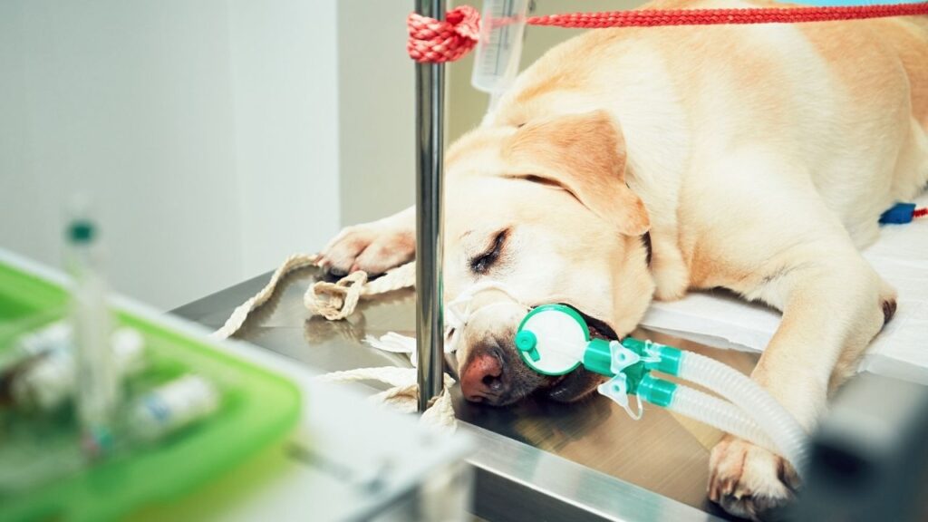
To obtain these images, your pet will need to lay still in a specific position (determined by what the veterinarian is trying to see) for a short period of time. Padded pillows are often used to make the position more comfortable, however, some positions may be uncomfortable or scary and your veterinarian may recommend sedation to help make the procedure easier for your pet.
Can I Be with My Dog During an X-Ray?
While safety regulations do not allow owners to accompany their pets while x-rays are taken, your dog will be attended to by caring technicians throughout the process.
Once your dog is in position and the x-ray machine has been turned on with the proper settings, a staff member will press a button to take the x-ray. With a whirr and a beep, x-rays will be produced and pass through the dog’s body to produce an image.
X-rays are completely painless, and your dog won’t feel anything. The noise that some machines make can be stressful for some dogs.
Sometimes your veterinarian may be able to give you an answer immediately after taking the x-ray, though other times these images may need to be sent off to a boarded radiologist for review.
Most veterinary facilities now have digital x-ray systems, allowing the images to be taken and processed very quickly. If your vet still has traditional x-rays, the image will need to be developed similarly to a film camera.
Reasons Your Dog May Need a Radiograph
Radiographs allow veterinarians to see deep into the body to assess bones, organs, and other soft tissues that may not be easy to evaluate through physical examination alone.
Because a radiograph produces a shadow-like image, it can sometimes be hard to see inside a hollow organ such as the stomach, intestines, or bladder. If a tumor is suspected in these locations, a technique called contrast radiography may be used which involves using special dye-like compounds that show up well on radiographs to highlight any potential tumors.1,2
Safety and Side Effects from Radiographs for Dogs
The radiation exposure from an x-ray is minimal. In fact, the American Cancer Society has estimated that a chest x-ray exposes an individual to no more radiation than they would encounter naturally over a ten-day period.3
Standard radiographs are unlikely to have any side effects. Contrast studies where the contrast material is ingested or swallowed may lead to gas or bloating. Contrast studies where the contrast is injected have been reported to sometimes cause itching or flushing of the skin in people.2
When Are Dog X-Rays Recommended?
Radiographs (x-rays) for dogs can be useful in many situations:
- Identifying primary tumors, particularly in the bones, abdomen, or chest
- Determining if a tumor has spread through the body to other organs, especially the lungs
- Diagnosing bone fractures
- Identifying an intestinal obstruction
- Evaluating the lungs
- Evaluating the shape and size of the heart
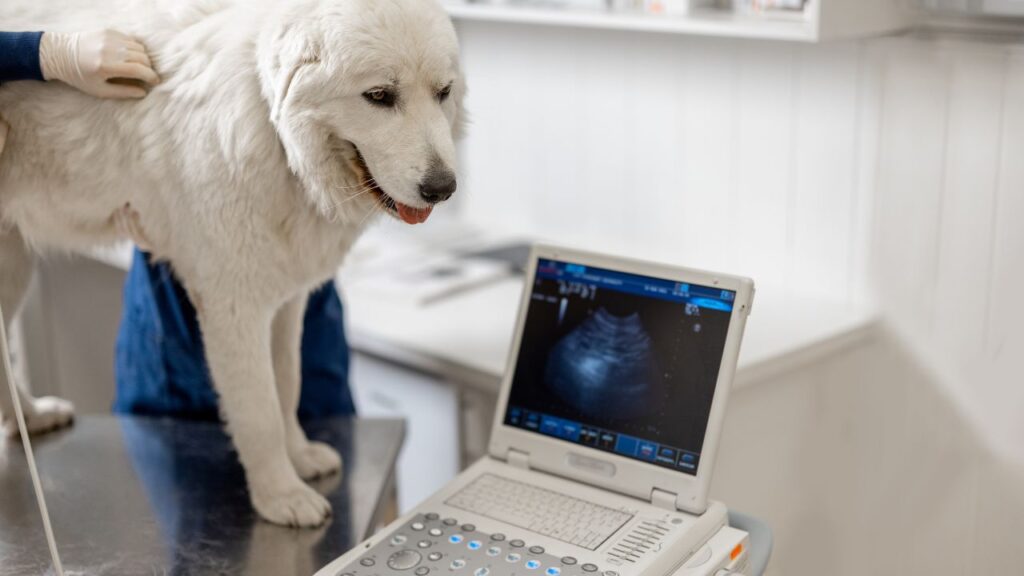
If your dog has cancer, radiographs are often combined with other imaging modalities such as ultrasound, CT, or MRI to get a complete idea of the extent of your pet’s cancer.
Cancers Radiographs Are Commonly Used For
Radiographs can be particularly helpful in diagnosing bone cancer as well as tumors in the abdomen or lungs.
They are also commonly used to determine if a tumor has metastasized, or spread, to the lungs.4
How to Get the Best Results from Dog X-Rays
Standard radiographs generally need no preparation.
However, if your pet is going to undergo sedation or have contrast administered, you may need to do some minimal preparation such as withholding food. Skipping breakfast can also be helpful to minimize the amount of food and poop inside your dog’s body. Ask your veterinarian if there is anything you need to do to prepare for your pet’s appointment.
Home Care
Most dogs do not need special home care after having x-rays.
If your pet was sedated for the procedure, they may have decreased energy level or appetite after their appointment. If contrast was given by mouth, you may notice discoloration of the stool (often white, from barium).2
Older pets, or those with arthritis, may be stiff or sore afterwards due to the specific positioning required to obtain a clear image.
Follow Up
Repeat radiographs may be necessary throughout your pet’s cancer treatment to help monitor any changes in the tumor.
Limitations of Radiographs (X-Rays) for Dogs
Some areas of the body are not easy to evaluate through radiographs, such as inside organs or in areas encased in bone (such as the skull).
For these locations a different type of imaging such as ultrasound, CT, or MRI may be more appropriate. Your veterinarian will make recommendations on the best type of imaging for your pet’s situation.
Where to Get a Dog X-Ray
Radiographs are readily available at most veterinary offices.
Depending on the situation, your veterinarian may be able to interpret the film for you immediately or it may need to be sent out for a specialist to review.
The Cost of Dog X-Rays
Many factors can influence the cost of radiographs including the area being imaged, whether your pet needs sedation, and whether the radiographs are being sent out to a radiologist for interpretation.
You can always ask your veterinarian for an estimate before any procedure so that you know the cost ahead of time. Pet insurance or payment options, such as CareCredit or Scratchpay, can help with the financial burden.
- UC Davis School of Veterinary Medicine. Small animal imaging: Radiography. https://www.vetmed.ucdavis.edu/index.php/hospital/diagnostic-imaging/small-animal/xray
- American Cancer Society. X-rays and other radiographic tests for cancer. https://www.cancer.org/treatment/understanding-your-diagnosis/tests/x-rays-and-other-radiographic-tests.html
- American Cancer Society. Understanding radiation risk from imaging tests. https://www.cancer.org/treatment/understanding-your-diagnosis/tests/understanding-radiation-risk-from-imaging-tests.html
- Suran JN. General Diagnostic Imaging in Small Animal Oncology. https://www.vet.upenn.edu/docs/default-source/penn-annual-conference/pac2016-proceedings/companion-animal-track-3/general-diagnostic-imaging–dr-suran.pdf
Topics
Did You Find This Helpful? Share It with Your Pack!
Use the buttons to share what you learned on social media, download a PDF, print this out, or email it to your veterinarian.

