EPISODE 217 | RELEASED May 22, 2023
How Does Ultrasound for Dogs Work? | Dr. Adrienne Anderson
Dog ultrasound is one of the many tools used to help diagnose cancer. Best of all? It’s painless!
SHOW NOTES
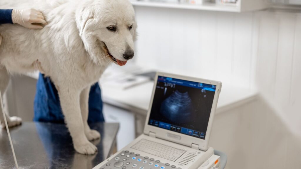
Veterinarian Adrienne Anderson explains how ultrasound for dogs works, when it is used, and where you can get it done with your dog. This “stellar diagnostic” lets your vet see what is going on inside your dog’s abdomen and heart in real time, without requiring sedation or surgery.
Many vet hospitals have their own basic ultrasound machine, or you can take your dog to a specialist to get a more thorough exam.
Listen in for all of the details on dog ultrasounds, as well as how this technology can be used to take biopsy samples.
Links Mentioned in Today’s Show:
Ultrasound Examination in Dogs article: https://www.dogcancer.com/articles/diagnosis-and-medical-procedures/ultrasound-examination-in-dogs/
[00:00:00] >> Dr. Adrienne Anderson: I would say it is generally one of the things that has the least cons about it in terms of diagnostics that we recommend because it is so non-invasive, it provides so much information, basically in immediate time, right, like you do it and that, you get the answers right then.
[00:00:19] >> Announcer: Welcome to Dog Cancer Answers, where we help you help your dog with cancer.
[00:00:25] >> Molly Jacobson: Hello, friend. I’m Molly Jacobson and today on Dog Cancer Answers, we’re looking at one of the diagnostic tools in your veterinarian’s toolbox: ultrasound. Ultrasound is available in specialty hospitals, but also many regular veterinary offices. And it’s a painless way to see what’s happening in your dog’s abdomen and some other places in the body.
To give us more details on how ultrasound works and when it’s useful for our dogs, we are joined by Dr. Adrienne Anderson. She’s a veterinarian from Colorado who has been writing amazing articles for our website, dogcancer.com. Dr. Adrienne Anderson, thank you so much for joining us today.
[00:01:06] >> Dr. Adrienne Anderson: My pleasure. I’m happy to be here.
[00:01:08] >> Molly Jacobson: Yeah, you’ve been doing such wonderful work with us on dogcancer.com and I just appreciate your love and attention.
[00:01:17] >> Dr. Adrienne Anderson: Yes, indeed.
[00:01:19] >> Molly Jacobson: So I think ultrasound is one of those things we all think we’re familiar with because we’ve encountered it in so many television and movies, right? Like-
[00:01:27] >> Dr. Adrienne Anderson: Yeah.
[00:01:27] >> Molly Jacobson: Pregnant woman gets ultrasound, people in our lives get ultrasound, we get ultrasound. So, but what is ultrasound?
[00:01:35] >> Dr. Adrienne Anderson: So ultrasound is a technology that is used to create images of the inside of the body, I guess is kind of how I would describe it in its simplest way. It’s a way of using sound waves in a non-invasive and non-painful way to make pictures of things that we’re really interested in looking at. So.
[00:01:57] >> Molly Jacobson: That’s so interesting that you can use sound waves to look.
[00:02:01] >> Dr. Adrienne Anderson: Yeah.
[00:02:01] >> Molly Jacobson: It’s counterintuitive.
[00:02:03] >> Dr. Adrienne Anderson: It’s amazing.
[00:02:03] >> Molly Jacobson: Yeah. I recently spoke to Dr. Joanne Tuohy, who is running these experiments on a high intensity focused ultrasound where they’re destroying bone tumors with sound waves.
[00:02:14] >> Dr. Adrienne Anderson: Yeah.
[00:02:15] >> Molly Jacobson: Which is very cool, but very different than what we’re talking about here.
[00:02:19] >> Dr. Adrienne Anderson: Totally. Yeah.
[00:02:20] >> Molly Jacobson: But.
[00:02:20] >> Dr. Adrienne Anderson: Fascinating.
[00:02:21] >> Molly Jacobson: Yes.
[00:02:21] >> Dr. Adrienne Anderson: And expanding our use of the technology, so.
[00:02:24] >> Molly Jacobson: Yeah. Yeah. It’s so interesting. Okay, so basically this technology is generating sound waves outside the body that enter the body and then kind of work with each other, and there’s some sort of a algorithmic, um, experience on a computer that takes the information the sound waves are interacting with and makes a picture.
[00:02:45] >> Dr. Adrienne Anderson: It’s essentially like, you know, a probe that sends signals through tissues in the body, whether that’s organ tissues or fluid. And that sound interacts with those tissues depending on what they look like differently, um, in terms of how dense they are, what shape they are, how thick they are, and then depending on that, sends back a signal. And how much is absorbed or reflected from that tissue or fluid provides a black and white or gray scale image of what that looks like.
And so we can tell the difference between, again, between fluid, between tissue, between tissue that’s different thickness or has a different texture or quality, uh, we can tell the difference between those things and that really helps us determine if something is wrong, really, or if something is normal or if there’s something we need to sample or, yeah.
[00:03:34] >> Molly Jacobson: What kinds of things can you see with ultrasound versus other imaging techniques like x-rays or MRI or CAT scan or?
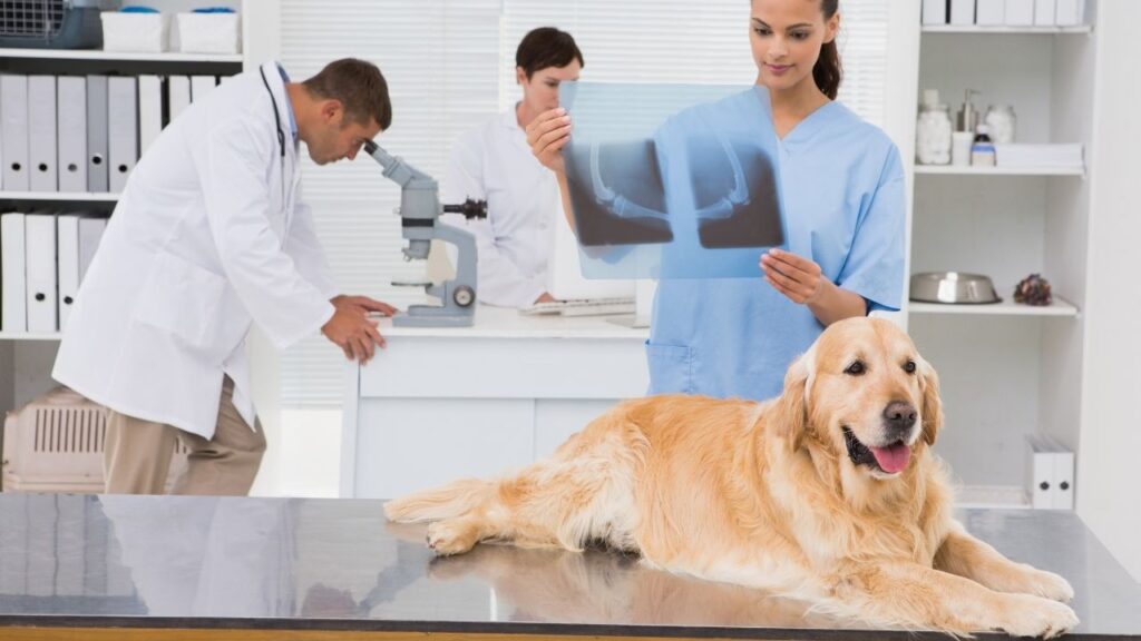
[00:03:42] >> Dr. Adrienne Anderson: Yeah, so ultrasound is, I guess if I’m comparing it first to x-ray, x-ray is really a method of creating a two-dimensional image of a three-dimensional space, which has value, and it’s really good for looking at bones, it’s really good at looking for things that have any mineral quality. There are certain things like in the chest that it’s really good at looking at, but it’s limited in its ability to create a three dimensional image. And x-ray is not super good at certain detail of the abdomen. It’s not great at giving us a lot of information for that.
[00:04:16] >> Molly Jacobson: It works better with harder surfaces rather than the soft, slippery organ surfaces.
[00:04:20] >> Dr. Adrienne Anderson: Exactly, exactly. Um, and ultrasound is really good at creating detail of the abdomen, I think is probably what we use it most for, although the heart as well, it, it does a really good job of creating a kind of a three-dimensional structure in a manner of speaking. And so it is better at getting detail for those types of structures. It’s not very good at nervous tissue, so it’s not very good at – ultrasound is not very good at looking at the brain or looking at the spinal cord.
We more use MRI for that, or in some cases, CT, although CT also kind of better for bone. But it is really good at looking in the abdomen and finding fluid in places that maybe it shouldn’t be, soft tissue structures, so you can look at tendons, you can look at some muscle structures and that sort of thing, but I would say that’s less frequently used in small animal than an equine practice, for example.
[00:05:16] >> Molly Jacobson: Okay, so bigger animal, you can maybe visualize bigger soft tissues, but-
[00:05:21] >> Dr. Adrienne Anderson: Yeah.
[00:05:22] >> Molly Jacobson: Smaller animal, we’re looking mostly at organs in the abdomen, it sounds like.
[00:05:26] >> Dr. Adrienne Anderson: You got it, mostly.
[00:05:27] >> Molly Jacobson: Okay.
[00:05:28] >> Dr. Adrienne Anderson: We sometimes will look at if there’s fluid in the sac that surrounds the lungs. It’s pretty good at looking at that, and finding that, and there’s some other applications for the chest, but I would say in small animal, we by far mostly use it for the abdomen and the heart.
[00:05:42] >> Molly Jacobson: Okay, so walk us through a typical ultrasound appointment. What actually happens and does it hurt? This is something that I get questioned about, like, does ultrasound hurt?
[00:05:52] >> Dr. Adrienne Anderson: It’s a great question. Ultrasound does not hurt in itself. It can be an anxiety provoking event for some animals or can increase their stress response a little bit, but the procedure itself is not painful. Kind of from start to finish, I suppose you, you typically will make an appointment, so you will book an appointment for – if you’re doing a formal ultrasound, a boarded radiologist most commonly does it, although there are general practice veterinarians that can perform some version of it. But a true formal ultrasound is scheduled in advance, typically.
[00:06:25] >> Molly Jacobson: Okay.
[00:06:25] >> Dr. Adrienne Anderson: And so depending on where you are, you may be given some medication to give by mouth if your dog or cat is very stressed, just to kind of make them a little bit less nervous. Or if they’re really low key and not stressed, will tolerate it well, then they can just do it without.
[00:06:44] >> Molly Jacobson: Okay.
[00:06:45] >> Dr. Adrienne Anderson: Some of them require an injectable sedation, so in that case there are medications that we can give as an injection to just calm them. And that is really twofold. Yes it calms them and by virtue of calming them gives the radiologist better images, or that gives the doctor much better images, um, that are more accurate.
[00:07:03] >> Molly Jacobson: They’re not squirming around and ruining the picture.
[00:07:06] >> Dr. Adrienne Anderson: Yeah, exactly. They’re not squirming around, which means they are feeling better and you are getting more information. And so those things together are advantageous, but that’s typically the first part. And then it’s really shaving of the fur. So wherever it is that the point of interest is, which I guess I’ll use the example of the abdomen in this case, they’ll shave a pretty wide margin around the abdomen.
And that’s really because the fur interferes with how those sound waves travel. And so better images are acquired when there is not fur in the way.
[00:07:37] >> Molly Jacobson: Okay.
[00:07:38] >> Dr. Adrienne Anderson: So then after that, it’s usually laying them down, either, often on, all the way on their back, which sometimes a little support boat is provided so that they’re more comfortable on their back, and then some radiologists and some clinicians will place them on their sides depending on what type of images. They’ll usually rotate throughout to get whatever views of those, to make those images three-dimensional and make sure that we have all the information that we can get.
And then it’s really a matter of a little probe, so I don’t know, I guess three to five inches across, essentially, that is covered in gel. So some sort of gel that helps to transmit those waves through the tissue. So either gel or alcohol, isopropyl alcohol, directly on the patient. Or gel on the probe itself. Sometimes both.
[00:08:28] >> Molly Jacobson: Okay.
[00:08:29] >> Dr. Adrienne Anderson: And then that probe is placed on the area of interest and moved around with pretty gentle pressure typically. And once they’re rotated and the images from all sides are acquired, we can get video of that. They can get still pictures of that. They can get something called Doppler information, so information about how blood is flowing through these various tissues and whether that’s normal or if they’re using ultrasound to guide another procedure then Doppler can be helpful with that so you know where all the vessels are. And then they just kind of stand up and go about their day.
[00:09:04] >> Molly Jacobson: Because it doesn’t require anesthesia and it doesn’t hurt. And so when-
[00:09:08] >> Dr. Adrienne Anderson: Exactly.
[00:09:09] >> Molly Jacobson: When sort of the indignity of being put on your back and slimed up and something waved around on your body, you’re gonna go, okay, whatever that was.
[00:09:16] >> Dr. Adrienne Anderson: Yeah. Exactly. And then the gel is wiped off and they’re typically, if they’re sedated, obviously if it’s something reversible, then that will be reversed. Or they’ll just kind of rest and go back to their normal selves once they’ve recovered from that mild bit of sedation. But it is an exceptionally rewarding procedure for how non-invasive it is.
[00:09:37] >> Molly Jacobson: Hmm. Meaning you get a lot of value out of it without having to put the dog through a lot.
[00:09:43] >> Dr. Adrienne Anderson: Yep. You get a whole lot of value out of something that doesn’t require anesthesia and doesn’t require really a whole lot of pre-thought other than fasting is a big part of it. So that’s the only part that I forgot to mention earlier, that it is ideal for a patient to be fasted or not have eaten the 12 hours prior. So usually they can have dinner the night before, but no breakfast the morning of. It gives-
[00:10:09] >> Molly Jacobson: Okay.
[00:10:09] >> Dr. Adrienne Anderson: -again, much better images. Yeah.
[00:10:11] >> Molly Jacobson: Meaning you’re not looking at food.
[00:10:13] >> Dr. Adrienne Anderson: You’re not looking at food, there’s less likely gas, which is really can kind of toggle the image and cover up things that should be visible. And differentiating between food and other things can actually be kind of hard ’cause sometimes they reflect waves in similar ways and so it can obstruct or make things that might be something we wanna identify harder to identify when there’s food, especially if we’re looking at the small intestine or the large intestines, somewhere in the gut where we really are interested. Food can obscure or make that much harder to see.
[00:10:43] >> Molly Jacobson: That makes sense. It can look like other things.
[00:10:45] >> Dr. Adrienne Anderson: Yeah.
[00:10:46] >> Molly Jacobson: And you can’t, you’re not getting like information about color, it’s all black and white, so you have to kinda go with shades of gray.
[00:10:53] >> Dr. Adrienne Anderson: Shades of gray.
[00:10:54] >> Molly Jacobson: Yeah.
[00:10:54] >> Dr. Adrienne Anderson: And sometimes food and and tissue, or at least food and foreign material, if that’s relevant, can be very similar looking and can confuse the value of what you’re trying to do.
[00:11:06] >> Molly Jacobson: Right. You mentioned that sometimes it’s used to guide a procedure. Can you talk a little bit about that?
[00:11:14] >> Dr. Adrienne Anderson: Yeah, so it is a real life, you know, live version of what’s happening in the abdomen. So if they’re doing the ultrasound exam and they say, oh wow, this, you know, looks like maybe there’s a tumor or some abnormal structure on the liver, for example. Then they can use the ultrasound to guide a picture of where, live, they are going to stick a needle, for example, to sample that area. They wanna make sure not only that they’re getting the right location, but also that we’re avoiding structures that we really don’t want to touch with a needle.
[00:11:48] >> Molly Jacobson: Right.
[00:11:48] >> Dr. Adrienne Anderson: So making sure that it’s the safest that it can be and the most accurate that it can be for sampling either tissue, so true tissue, like a tumor or something, or fluid. So if we know there’s fluid hanging around in the abdomen, it’s a really good way to accurately get to the right place to get a sample of it.
[00:12:07] >> Molly Jacobson: Ah, so like, yeah, if, if you’ve got a packet of fluid somewhere and, where it shouldn’t be, you can use the ultrasound, right?
[00:12:14] >> Dr. Adrienne Anderson: Yep.
[00:12:14] >> Molly Jacobson: Do they typically do that during the ultrasound that’s been prescheduled and just go in there right away?
[00:12:19] >> Dr. Adrienne Anderson: Usually.
[00:12:20] >> Molly Jacobson: And say, yeah, usually. Okay.
[00:12:21] >> Dr. Adrienne Anderson: Usually. They typically will, for that type of sampling, ensure that sedation is a part of it because that is a very sensitive procedure. You wanna make sure the patient is still, because if they move, it’s more dangerous.
[00:12:35] >> Molly Jacobson: Right.
[00:12:35] >> Dr. Adrienne Anderson: So usually if they’re not already sedated, ’cause it’s a very compliant patient, often sedation will be administered if that’s gonna be a part of the deal.
[00:12:41] >> Molly Jacobson: I see. So your dog might not have gotten sedation for the ultrasound, but then if they see something, they’ll say, okay, now let’s give them some sedation so we can get a sample. Well, that makes sense.
[00:12:51] >> Dr. Adrienne Anderson: The caveat I guess, would be like, the urinary bladder. So if they’re gonna take a sample from, of urine directly from the bladder, ’cause it’s a sterile way to get urine, then often sedation is not necessary for that.
[00:13:04] >> Molly Jacobson: Oh really? Okay.
[00:13:06] >> Dr. Adrienne Anderson: No.
[00:13:06] >> Molly Jacobson: Huh. It just is accessible enough from the outside that you kinda, the bladder’s just sitting right there.
[00:13:12] >> Dr. Adrienne Anderson: It’s sitting in a good place that it really doesn’t have a whole lot that’s right above it. I guess if you’re, the patient’s on its back, and this is the surface of the abdomen-
[00:13:22] >> Molly Jacobson: Uh-huh.
[00:13:22] >> Dr. Adrienne Anderson: -the bladder is pretty close to the surface. And so sampling it that way is actually not as sensitive geographically, if you will, as other places in the abdomen.
[00:13:35] >> Molly Jacobson: Okay, ’cause it’s not too deep in the dogs, like dogs tolerate needles pretty well when they’re relaxed, right?
[00:13:41] >> Dr. Adrienne Anderson: They do.
[00:13:41] >> Molly Jacobson: Yeah.
[00:13:41] >> Dr. Adrienne Anderson: Exactly.
[00:13:42] >> Molly Jacobson: Yeah. They don’t look at them necessarily and go, oh my God, like we do.
[00:13:45] >> Dr. Adrienne Anderson: Oh my God. Yeah, some of them do, but most of the time, most of the time, they are pretty tolerant of that.
[00:13:54] >> Molly Jacobson: Okay, well that’s good to hear. We’re going to take a short break, hear from our sponsors, and then come back with more information on ultrasounds for dogs.
And we’re back with Dr. Adrienne Anderson. So when would a veterinarian usually recommend getting an ultrasound? And how common would you say it is for you to recommend an ultrasound for a patient?
[00:14:21] >> Dr. Adrienne Anderson: I would say it is very common. I probably in a week recommend it three to five times, I would say.
[00:14:31] >> Molly Jacobson: Okay. And I live in a sort of very specific, you know, part of the planet. Maui is incredibly isolated in general, and we get used to things just not being here. We’re just like, oh, we just can’t get that here.
[00:14:43] >> Dr. Adrienne Anderson: We just can’t get it. Yeah.
[00:14:44] >> Molly Jacobson: Yeah. So there is a veterinarian who I don’t think is boarded, but she comes around and she does ultrasound and like she’s at a practice one day and then another practice the next day, and-
[00:14:54] >> Dr. Adrienne Anderson: Yeah.
[00:14:54] >> Molly Jacobson: We have to kind of plan these things carefully. So I think of ultrasound as being something that’s sort of not really commonly available.
[00:15:02] >> Dr. Adrienne Anderson: Right.
[00:15:02] >> Molly Jacobson: But I live in a pretty rural isolated place. So how common is it? Like where can you get ultrasound in general, would you say?
[00:15:09] >> Dr. Adrienne Anderson: That is a great question. I happen to be in an area with a high density of specialty medicine.
[00:15:16] >> Molly Jacobson: You’re in Colorado.
[00:15:16] >> Dr. Adrienne Anderson: And because of that – yeah.
[00:15:18] >> Molly Jacobson: Yeah.
[00:15:18] >> Dr. Adrienne Anderson: In the front range of Colorado where there is a lot of specialty medicine. And so we do, at the clinic that I work in, use a boarded radiologist service with multiple radiologists that travel throughout our area and they happen to be very readily available. Often we can call them and get a same day image scheduled.
[00:15:40] >> Molly Jacobson: Oh, that’s nice.
[00:15:40] >> Dr. Adrienne Anderson: But that is by far not the case for many places. And I would say usually you’re looking at a few days, or if you’re doing something travel wise and it’s available but not in high volume, then either a longer delay or there are emergency facilities and some general practice facilities that have their own ultrasound machines, and many of them have extra training in their ability to measure and see structures and evaluate the meaning of, of those findings. And certainly that is becoming more common because access to specialty medicine is just not always so easy.
[00:16:19] >> Molly Jacobson: Right. Do you consider ultrasound a specialty diagnostic?
[00:16:24] >> Dr. Adrienne Anderson: Um, I think I don’t, in the sense that it is very useful even in a general practice setting. So a general practice veterinarian can use it to look for large problems. So look for is there fluid where there shouldn’t be fluid? Is there a large tumor that I can see pretty readily? There are systems for doing screening like that to look for major things.
But when it comes to smaller detail, measuring certain gland structures or you know, the thickness of the gastrointestinal wall, or, there are certain things that are more specialized and more challenging, I would say for someone who doesn’t have the experience of being boarded in radiology.
[00:17:08] >> Molly Jacobson: Okay.
[00:17:08] >> Dr. Adrienne Anderson: But I wouldn’t say that it itself is a specialty service ’cause it is so useful for, for simpler things in general practice as well.
[00:17:17] >> Molly Jacobson: Seeing pennies in the, in the obstruction.
[00:17:20] >> Dr. Adrienne Anderson: Yes.
[00:17:20] >> Molly Jacobson: Oh.
[00:17:20] >> Dr. Adrienne Anderson: Yes. Or sometimes using it in general prac, I would say a lot of general practice uses it for what we were talking about before, taking urine from the bladder. You can know anatomically where the bladder is and many places do what we call a blind stick. So if you need a sterile sample of the bladder, you find the bladder, you either feel it or you know pretty much where it is, and you put the needle in and you get urine. A lot of places do that.
But if you have an ultrasound, you can look at the bladder and know exactly where your needle’s gonna go. And that is a safer procedure. And I think something that most veterinarians would prefer to have if they’re doing something like that. Yeah.
[00:17:58] >> Molly Jacobson: So I’m imagining that it’s a useful tool, but many practices don’t have, it must be overhead, just cost of the machine.
[00:18:07] >> Dr. Adrienne Anderson: The machine is expensive usually, I mean, it depends on the quality of the machine, but for a very nice one, they’re usually tens of thousands, in the low tens of thousands to get a high quality one that is really producing images that you can measure and get detail of texture and that sort of thing. I think you can get ’em for under $10k, but that’s really ones you’re using like for what we were talking about, getting urine from the bladder. I can see the bladder, I know where it is.
I can see that there’s a lot of fluid in this area and I want to take it out. So things that don’t require intense detail, but allow you to see the difference between a fluid and a tissue.
[00:18:43] >> Molly Jacobson: Okay, I see. So there’s different range of machines and they have different-
[00:18:47] >> Dr. Adrienne Anderson: Yeah.
[00:18:47] >> Molly Jacobson: -capacities and the more detailed ones are gonna allow, like I would imagine, I got a echocardiogram a couple years ago and she was measuring absolutely everything right on the screen. So that’s what you’re talking about.
[00:19:01] >> Dr. Adrienne Anderson: Yeah.
[00:19:02] >> Molly Jacobson: That higher end ultrasound machine is allowing you to do all sorts of things that a lower quality one would not. The technology’s the same.
[00:19:10] >> Dr. Adrienne Anderson: Yeah, the technology’s the same. But yeah, like echocardiogram is a very good example of that because often the, like a boarded cardiologist who’s doing echocardiograms in veterinary medicine has a very high quality machine that includes Doppler, so the blood flow portion, which is by necessity requires a nicer machine and provides more detail.
[00:19:30] >> Molly Jacobson: Okay.
[00:19:30] >> Dr. Adrienne Anderson: And the heart is something that we, that that is used for, for sure.
[00:19:34] >> Molly Jacobson: Okay. So that is just something that a, a specialty center that’s gonna use it more often is gonna be more likely to have it.
[00:19:41] >> Dr. Adrienne Anderson: Yeah.
[00:19:41] >> Molly Jacobson: That makes sense. So how much does ultrasound generally cost? I mean, obviously there’s huge regional differences and if you’re the only practice with an ultrasound, I suppose you can charge what you want, but in general, what can people?
[00:19:55] >> Dr. Adrienne Anderson: Yeah, it’s a good question. So I would say usually between $300 and $600 or $700, depending on, does your animal need sedation, so is that a part of it.
[00:20:09] >> Molly Jacobson: Okay.
[00:20:10] >> Dr. Adrienne Anderson: Is a boarded radiologist performing it and viewing it? That is on the higher end of the cost spectrum, for sure. And how many that company or group, how many ultrasounds in a given period of time do they perform. And I would say those, those factor into what cost you’re looking at.
[00:20:28] >> Molly Jacobson: Okay. Even though we think of specialty medicine as more expensive, sometimes it’s less expensive when you consider that they can scale these costs.
[00:20:37] >> Dr. Adrienne Anderson: Yeah.
[00:20:37] >> Molly Jacobson: They can, yeah, they have access to the machines that they need, and so you don’t have to like go out of your way or have someone else come out of their way to come to you with that machine.
[00:20:47] >> Dr. Adrienne Anderson: Yeah. Or even if, so here, for example, yes, we have a boarded radiologist that travels and because he comes so often and he’s moving around all the time to do these images, like he doesn’t charge an emergency fee were I to call and say, Hey, can you come now? He’d be like, yep, I’ll be there in a couple hours. Whereas in a lower volume area, they may charge a premium on rush service like that.
[00:21:11] >> Molly Jacobson: Right.
[00:21:11] >> Dr. Adrienne Anderson: So an emergency service.
[00:21:13] >> Molly Jacobson: Right. Right, right, right. Okay, great. Is there any time when you would be cautious about recommending ultrasound for a dog? Are there any cons to getting an ultrasound?
[00:21:25] >> Dr. Adrienne Anderson: I would say it is generally one of the things that has the least cons about it in terms of diagnostics that we recommend, because it is so non-invasive, it provides so much information, you know, basically an immediate time, right, like you do it and that, you get the answers right then.
So I think the only time that I’m hesitant is if there’s financial concern for a pet’s guardian where, you know, I would be choosing, am I gonna do this diagnostic or am I gonna treat this patient to be more comfortable? Or to, if there’s something that I can selectively improve quality of life for versus getting a diagnostic answer or doing an ultrasound to get an answer, then I probably would opt for, for something that will improve the patient’s life in the immediate, if I need to make that choice.
[00:22:14] >> Molly Jacobson: Right.
[00:22:14] >> Dr. Adrienne Anderson: So I think only in the sense that I am picking something over something else for a financial reason, in that context, would I say maybe don’t do it.
[00:22:24] >> Molly Jacobson: That makes sense. So it’s really completely safe and if, if you can afford it, it’s gonna give you more information to help you make treatment plans.
[00:22:32] >> Dr. Adrienne Anderson: Yeah.
[00:22:33] >> Molly Jacobson: Yeah.
[00:22:33] >> Dr. Adrienne Anderson: Yeah. I would say there are, in terms of like if you’re sampling something, so sampling the liver or sampling an organ, that I think comes with a separate set of risk that I would reevaluate that. So.
[00:22:45] >> Molly Jacobson: But it’s not from the sonogram. It’s not from the ultrasound.
[00:22:48] >> Dr. Adrienne Anderson: You’ve got it. It is not from the imaging itself or from the, from the ultrasound itself, so.
[00:22:53] >> Molly Jacobson: Right.
[00:22:53] >> Dr. Adrienne Anderson: It is thankfully so safe and gives so much information in such a short period of time that it is a stellar diagnostic.
[00:23:02] >> Molly Jacobson: Well, thank you so much for joining us to talk about ultrasound. I hope you’ll come back.
[00:23:07] >> Dr. Adrienne Anderson: Yeah, certainly.
[00:23:08] >> Molly Jacobson: And talk about other things with us.
[00:23:10] >> Dr. Adrienne Anderson: Excellent. I’d love to.
[00:23:12] >> Molly Jacobson: Okay.
And thank you for listening, friend.
So if an ultrasound is recommended for your dog, rest assured it’s quick, it’s painless, and it can often give an awful lot of useful information about what’s going on in your dog’s body. Depending on the exact machine your vet has, she may even be able to show you photos or videos of your dog’s ultrasound. To read Dr. Anderson’s article about ultrasound for dog cancer, visit our website dogcancer.com where you’ll find many, many other articles about all things dog cancer as well.
You’ll also find a link to that and others in our show notes. And don’t forget to subscribe to Dog Cancer Answers in your favorite podcast app so you won’t miss future explainer episodes like this one.
If your dog has had an ultrasound as part of their cancer journey, you can always share your story in our Facebook group. Find it at dogcancersupport.com, or just search for Dog Cancer Support in Facebook. It’s a wonderful community of dog lovers sharing their experiences and helping each other navigate this very difficult time.
I’m Molly Jacobson. From all of us here at Dog Podcast Network, I’d like to wish you and your dog a very warm, Aloha.
[00:24:32] >> Announcer: Thank you for listening to Dog Cancer Answers. If you’d like to connect, please visit our website at dogcancer.com or call our Listener Line at (808) 868-3200. And here’s a friendly reminder that you probably already know: this podcast is provided for informational and educational purposes only. It’s not meant to take the place of the advice you receive from your dog’s veterinarian.
Only veterinarians who examine your dog can give you veterinary advice or diagnose your dog’s medical condition. Your reliance on the information you hear on this podcast is solely at your own risk. If your dog has a specific health problem, contact your veterinarian. Also, please keep in mind that veterinary information can change rapidly, therefore, some information may be out of date.
Dog Cancer Answers is a presentation of Maui Media in association with Dog Podcast Network.
Hosted By
SUBSCRIBE ON YOUR FAVORITE PLATFORM
Topics
Editor's Picks
CATEGORY
